Safe • Accurate • Takes just 20 minutes
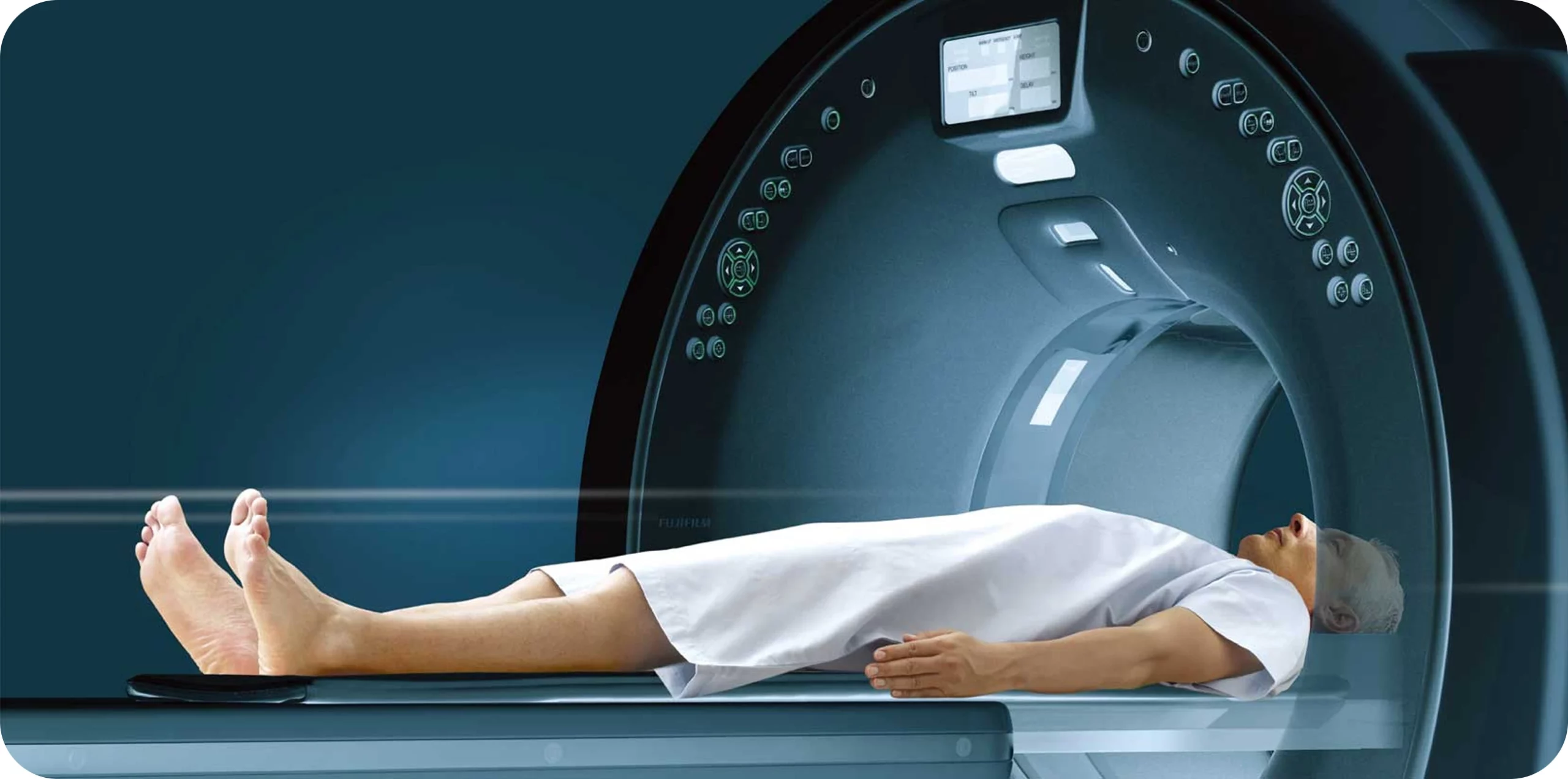
A computerized tomography scan, more commonly called a CT scan, allows doctors to see internal organs in much greater detail than a regular X-ray. It takes a series of X-ray images at various angles around your body and processes it together to create multiple cross-sectional images (slices) of the scanned body part.
Designed to ease your anxiety
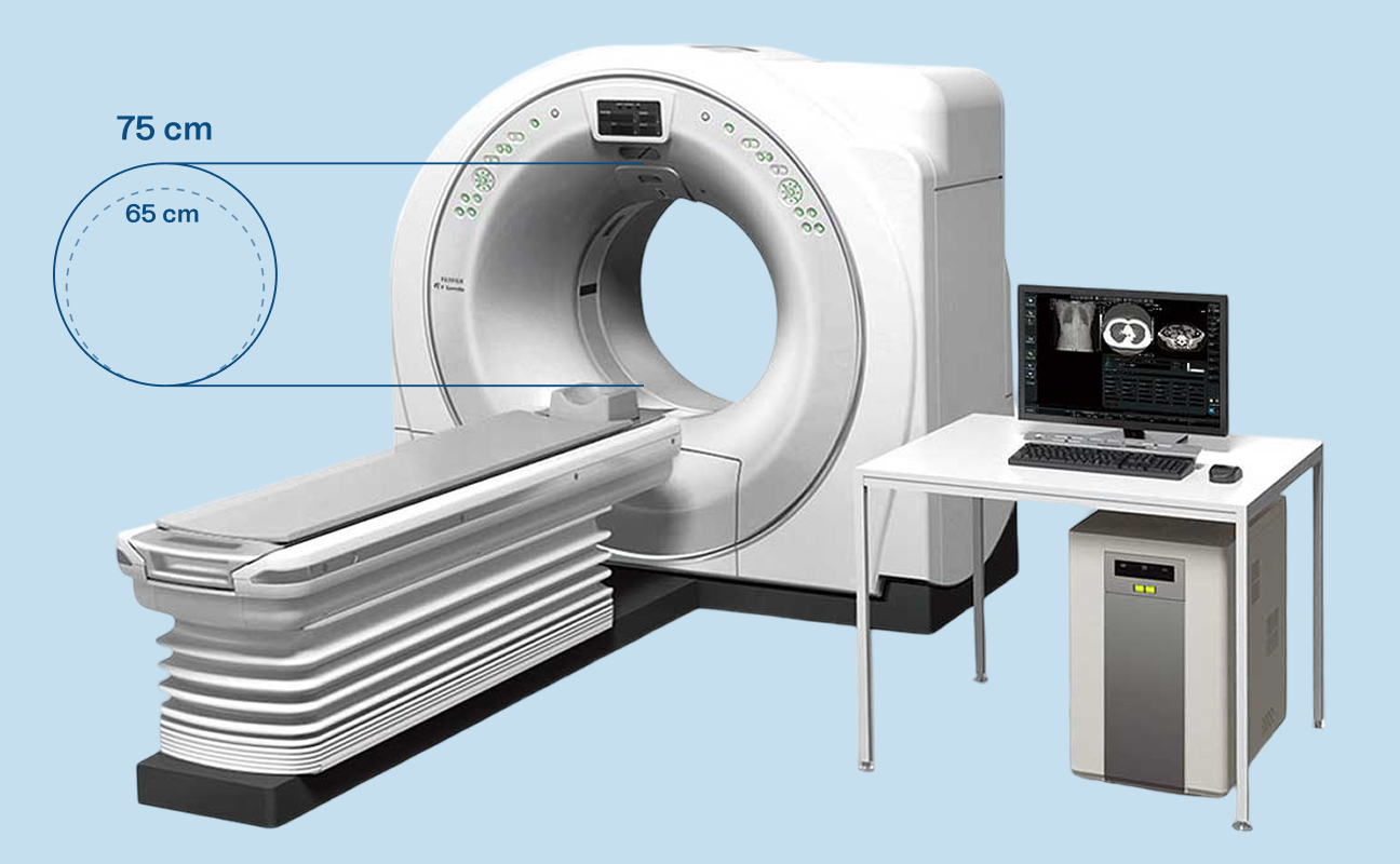
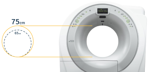
Our CT scan machines are designed to make the screening process as comfortable as possible. Our gantry bore (the opening in the CT machine) is larger than other machines to reduce the feeling of claustrophobia and ease your anxiety.
The accuracy of high-speed scanning
Our high speed scanning means you only need to hold your breath for 7-8 seconds. We use the latest 3D reconstruction technology that provides rapid coverage for an efficient and precise examination.
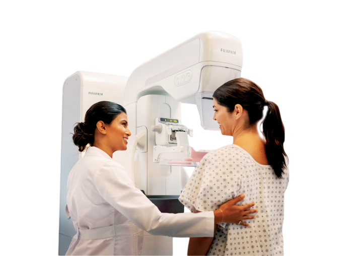
Minimum discomfort • Accurate • Takes just 10 minutes
Digital mammography is a screening test that takes scans of the breast tissues to assess breast health. Each breast is separately compressed against a highly sensitive digital flat panel to obtain the best quality images for examination.
Flexible paddle for added comfort
To make the screening process more comfortable, our digital mammography machines use a flexible compression paddle. This allows the machine to gently fit the natural curve of the breast, allowing the pressure to be dispersed evenly and lowering your discomfort.
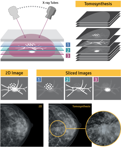
Accurate screening
We use 2D imaging, along with AI technology to comprehensively examine all sides of the breast.
During the screening process, the mammography machine moves in an arc around the breast to capture a series of x-ray images at different angles.
The images are intelligently reconstructed that allows doctors to make quick and accurate diagnoses. The images make it easier to identify abnormalities when compared to regular mammography, where the overlapping breast structures makes it harder to identify an abnormality.
Comfortable • Accurate • Takes just 20 minutes
Endoscopy is used to examine a person’s digestive tract. An endoscope is a thin, flexible tube with a small light and camera on the end. It allows doctors to view high quality images of the digestive tract on a monitor and observe for abnormalities.
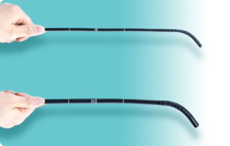
Ultra-Slim Design
Our ultra-slim nasal endoscope enables our screening process to be virtually painless and less nauseating than a traditional oral endoscope. It allows us to screen you without the use of sedatives, making the process safer.
Cutting-edge Multi Light technology
Our Multi Light™ technology creates high-quality images with observation modes that allow our doctors to make better and more accurate diagnoses.
White Light Imaging provides sharper and brighter images.
Linked Colour Imaging (LCI) gives us increased colour contrast that helps in detecting lesions or inflammation.
Blue Light Imaging (BLI) give us an improved and accurate contrast imaging.